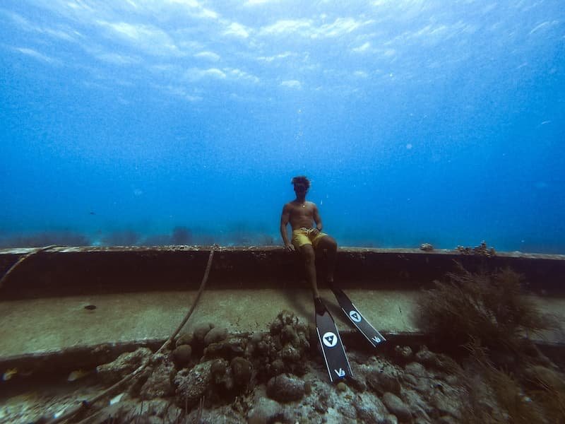
 Nick Pelios
Freediver, Creator
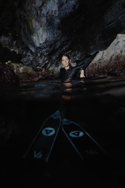
Nick Pelios
Freediver, Creator
Share this on
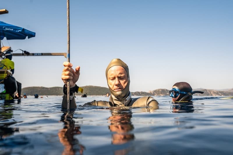
Nick Pelios
Freediver, Creator


When freedivers describe “flow at depth,” they often mean a state of absolute mental quiet, acute awareness, and seamless integration of body and mind under extreme physiological stress. The neurology behind that state is an intriguing frontier: how much does it overlap with the classical “flow” of athletic performance, or the meditative states of contemplative practice? In this essay I explore and compare EEG signatures of static apnea (dry breath hold), meditation, and high-intensity flow in extreme sports, drawing on empirical studies and case reports. The goal is to sketch a neurophysiological model of “flow at depth,” highlighting similarities, divergences, and gaps in our current knowledge.
In cognitive neuroscience, the flow state is typically defined as a state of immersive engagement, where subjective time may distort and action feels effortless. In EEG studies of flow, a recurring pattern is increased theta power in frontal regions, paired with moderate increases in alpha in frontal and central cortex. Katahira et al. (2018) found that during flow, frontal theta amplitude was elevated relative to baseline, and frontal or central alpha also rose moderately. In these contexts, theta is often associated with internal attention, error monitoring, and resource allocation; alpha is thought to signal suppression of task-irrelevant networks or cortical idling in nonessential areas.
Other investigations explore event related potentials. Yun et al. (2017) used suppression of auditory evoked potentials as a passive probe: deeper flow corresponded with greater suppression of auditory responses, interpreted as gating of external sensory input in favor of task immersion. Their analysis also implicated increased beta activity and connectivity in the anterior cingulate cortex and temporal poles. This hints that flow is not simply quiet mind but an actively modulated state of attentional control and network gating.
A recent preprint called Quantifying Flow State Dynamics (2025) used a prefrontal EEG device alongside subjective questionnaires in athletic tasks. The authors reported significant alignment between EEG metrics in prefrontal theta and alpha dynamics and subjective reports of flow, supporting the feasibility of real-time flow detection from frontal cortex signals. While such approaches remain preliminary, they reinforce the view that frontal theta and alpha interplay is central to flow.
Thus, in many flow paradigms, one sees an inverted U of cortical engagement: a boost in theta to sustain internal focus and cognitive control, together with alpha-mediated suppression of irrelevant input. The reconfiguration may also involve coherence, phase synchrony, or connectivity shifts across attentional and default mode networks, but to date those network dynamics are less thoroughly quantified in flow research.

Meditation research offers more extensive EEG literature, albeit with wide heterogeneity across styles. A consistent theme is slowing down of cortical rhythms: increases in theta (4–8 Hz), particularly midline frontal theta, and increases in alpha (8–12 Hz), often in posterior or parietal regions, sometimes with elevated coherence or synchrony. For instance, Ahani et al. (2014) studied mindfulness meditation in novices: after a six week intervention, EEG spectral differences in theta, alpha, and respiration coupling allowed discrimination of meditative versus control states with around 85 percent accuracy.
Meta reviews point out caveats: different meditation styles, subject expertise, signal processing choices, electrode montages, and baseline comparisons complicate generalization. One careful review (Deolindo et al. 2020) emphasizes that some reported effects might be artifacts of methodological variability rather than robust neurophysiological signatures.
Still, some recurring motifs emerge: experienced meditators may show greater theta and alpha increases, reduced beta, and enhanced synchronization in low frequencies. Some studies also report increases in gamma or cross frequency coupling during deep meditation, perhaps signaling integration or insight states.
Importantly, meditation is generally a low metabolic, low arousal state. Oxygen consumption, heart rate, and sympathetic activity are lower than during strenuous tasks. The neurophysiology involves sustained attentional focus or nonjudgmental awareness, but under conditions of metabolic ease. That contrasts sharply with the enforced constraints of static apnea, where the brain must operate under progressively worsening hypoxia and hypercapnia.
In the freediving domain, EEG and complementary imaging is still emergent. A landmark case study (Annen et al. 2021) examined a world champion freediver performing prolonged static dry apnea using high density EEG and fMRI. Compared to baseline rest, the apnea state showed increased EEG power and functional connectivity in the alpha band, along with decreased delta connectivity. The authors interpret this as evidence that the diver enters a mental state overlapping with meditative and attentional signatures but also retains unique features attributable to apnea stress.
Specifically, during apnea the global alpha connectivity rose from baseline mean connectivity 0.024 to 0.045, a statistically significant increase. Regionally, the diver showed reduced delta connectivity especially between frontal and occipital, and temporal parietal nodes. In fMRI, connectivity within the Default Mode Network increased, and visual network connectivity increased, whereas sensorimotor areas lost connectivity.
That report also connects phenomenology: the diver described a functional dissociation between body sensations and mental focus, almost as if the mind creates a bubble to isolate itself from peripheral discomfort. The shift in connectivity patterns might reflect this suppression of somatosensory integration while reinforcing internal networks.
Another relevant study measured total EEG spectral amplitude across resting, rhythmic breathing, and apnea phases. They did not find large spectral shifts compared to baseline resting, at least in median amplitude values for the 0.5–30 Hz band, though subtle interval changes emerged. Their data suggests that gross EEG amplitude might remain stable in some breath holders, especially in early phases of apnea, but effects may be more in connectivity or regional distribution rather than amplitude.
Interpreting these findings is delicate. One must keep in mind the confounds of hypoxia, hypercapnia, vascular constriction, and changes in skull conductivity or CSF thickness under altered blood flow. The instantaneous jump in alpha connectivity suggests the effect is not simply accumulating hypoxic damage but an active reconfiguration of network gating in response to breath holding.
To bring these threads together, imagine three overlapping regimes on a spectrum of immersion: meditation with low metabolic stress and sustained attention, sport flow with high activation and rapid feedback loops, and static apnea as a hybrid of stress and inward attention. The neural correlates seem to share a core domain of theta and alpha modulation and network gating, albeit adapted to each context’s constraints.
In classical flow, frontal theta boosts internal cognitive control, while alpha suppresses irrelevant networks. In meditation, theta and alpha shifts emerge under low arousal, encouraging introspection and cortical quieting. In static apnea, the diver’s brain appears to amplify alpha connectivity and reduce delta coupling, perhaps as a protective strategy: suppress peripheral sensory noise, reinforce Default Mode and attentional loops, and maintain attentional equilibrium under metabolic stress.
One speculative framework is that during apnea, the brain enters a constrained flow. It is simultaneously managing physiological threat from rising carbon dioxide and falling oxygen and preserving focused awareness. The increase in alpha connectivity may act as a shield against distracting perceptual noise; the reduction in delta connectivity may reflect disconnection from baseline sensory integration. In effect, the diver’s mind constructs a tunnel of internal coherence.
Yet there are qualitative differences: in sport flow, the pace is dynamic and motor sensory integration is central. In apnea, the body is largely quiet and involuntary autonomic systems dominate. In meditation, the body is at ease and minimal external feedback guides attention. So while the shared EEG motifs reflect common design, the divergences tell us that each flow state is tuned to its domain.
Importantly, the apnea literature is extremely limited. The case study of one elite diver is suggestive but not generalizable. We lack group studies of EEG during maximal apnea, comparisons across freedivers of different expertise, or time resolved dynamics of spectral transitions during descent and ascent of apnea. We also know little about cross-frequency coupling, coherence gradients, or directionality of connectivity under apnea stress.
Understanding the neurology of flow at depth is a bold frontier. The emerging data imply that static apnea nudges the brain into a state that shares core features with both meditation and sport flow: enhanced internal coherence, network gating, and suppression of external noise. But it also imposes unique constraints of hypoxia, hypercapnia, and autonomic remodeling.
To evolve this into a robust model, future research should aim for group experiments with trained versus novice apnea practitioners, high density EEG plus source localization to isolate cortical generators, simultaneous measurement of physiological parameters such as oxygen saturation, carbon dioxide, heart rate, and cerebral oxygenation to correlate with neural shifts, and dynamic analysis of transitions across frequency bands and connectivity. Ideally such experiments could include control tasks: meditative rest, motor tasks inducing flow, and breath control without apnea, to better disentangle shared versus unique signatures.
If successful, this research could decode how elite freedivers sculpt their inner states under extreme constraint, and perhaps reveal principles applicable to other domains of human performance. The neurology of flow states at depth remains an invitation to chart a new map at the intersection of mind, body, and constraint.
References
Annen J, et al. Mapping the functional brain state of a world champion freediver in static dry apnea. Brain Structure and Function, 2021.
Katahira K, et al. EEG Correlates of the Flow State. PMC, 2018.
Yun K, Doh S, Carrus E, Wu D A, Shimojo S. Neural correlates of flow using auditory evoked potential suppression. Preprint, 2017.
Ahani A, Wahbeh H, Nezamfar H, et al. Quantitative change of EEG and respiration signals during mindfulness meditation. J NeuroEngineering Rehabilitation, 2014.
Deolindo CS, et al. A Critical Analysis on Characterizing the Meditation. Frontiers in Systems Neuroscience, 2020.
Prolonged dry apnoea: effects on brain activity and physiological functions. Ratmanova et al.
Rosso G, Ricci R, Pia L, Rebaudo G, Guindani M, Marocchino A, De Pieri G, Filippo Rosso A. Quantifying Flow State Dynamics: A Prefrontal Cortex EEG Based Model Validation Study. arXiv preprint, 2025.